
This section is The content in this section is supplied by GlobeNewswire for the purposes of distributing press releases on behalf of its clients. Postmedia has not reviewed the content. by GlobeNewswire Article content
September 3, 2026
Sign In or Create an Account
or View more offersArticle content
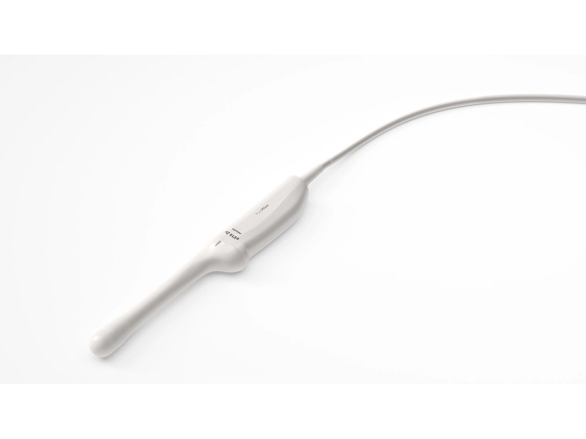
FDA 510(k) cleared and CE marked, the eV14-2v combines exceptional image quality and broad clinical capabilities in one lightweight transducer, helping streamline exams, support diagnostic confidence, and improve the patient experience
Article contentWe apologize, but this video has failed to load.Try refreshing your browser, or
tap here to see other videos from our team.Article content
Amsterdam, the Netherlands – Royal Philips (NYSE: PHG, AEX: PHIA), a global leader in health technology, today introduced the eV14-2v transvaginal transducer, expanding its obstetrics and gynecology ultrasound portfolio. FDA 510(k) cleared and CE marked, the eV14-2v is designed to support a broad range of gynecology and early obstetrics exams by combining image quality and broad clinical capabilities in a single lightweight transducer. The new transducer will be showcased at the 36th International Society of Ultrasound in Obstetrics and Gynecology (ISUOG) World Congress.
Article content
Story continues below
This advertisement has not loaded yet, but your article continues below.
Article content
Across obstetrics and gynecology, clinicians care for women with diverse clinical needs, from routine gynecologic exams to early pregnancy assessments and more complex cases. Achieving the desired balance of penetration and image resolution may require switching between transducers, which can interrupt workflow and extend exam times.
Article contentArticle content
The eV14-2v is designed to address these challenges by providing enhanced penetration, improved resolution, and a wider field of view in a lightweight, ergonomic design. The transducer delivers a 41.7% increase in penetration, a 10% increase in 2D resolution, a 15.8% improvement in field of view, and a 22% lighter design, supporting imaging across routine and complex gynecology and early obstetrics exams [1].
Article content
“Advancing women’s health is a priority for Philips ultrasound, and we are committed to developing innovations that make women’s healthcare more accessible and more comfortable for patients,” said Ajay Gannerkote, Ultrasound Business Leader at Philips. “At Philips, we’re focused on bringing together high-quality imaging, intelligent workflows and connected capabilities to simplify the ultrasound experience for clinicians and patients. The eV14-2v transducer reflects that approach, giving clinicians greater versatility in a single transducer and helping support high-quality care for women.”
Article content
Story continues below
This advertisement has not loaded yet, but your article continues below.
Article content
Built specifically for gynecology and obstetrics imaging, the eV14-2v leverages PureWave crystal technology, first introduced by Philips, and features a lightweight ergonomic design that supports user comfort during exams. This gives clinicians detailed visualization of anatomy relevant to evaluating infertility and pelvic pain associated with conditions such as endometriosis and fibroids. Philips technology, including 2D and color resolution, supports the detection of fetal abnormalities, with the ability to delineate the cardiac chambers in a fetus as early as 13 weeks when the heart is the size of a single corn kernel [2].
Article content
“Reducing transducer changes during exams has a meaningful impact, not just on efficiency, but on the patient experience as well,” said Dr. Michael S. Ruma, MD, MPH, Perinatal Associates of New Mexico. “With the eV14-2v, we’re able to complete more exams with a single transducer while maintaining excellent image quality, which helps streamline our workflow and improve patient comfort.”
Article content
The eV14-2v transducer can be used with Philips Collaboration Live, supporting real-time remote consultation during ultrasound exams. Clinicians can view live imaging, provide guidance during exams, and consult with care teams without requiring patient travel. This approach can help expand access to specialty care in underserved communities while supporting more efficient care delivery.